
Hot off the press: published paper in Small!
The collaborative work between the N&N group and the Eye Group from the University of Tampere (Finland) has finally materialized in common publication merging biomaterials and stem cell expertise!

The collaborative work between the N&N group and the Eye Group from the University of Tampere (Finland) has finally materialized in common publication merging biomaterials and stem cell expertise!

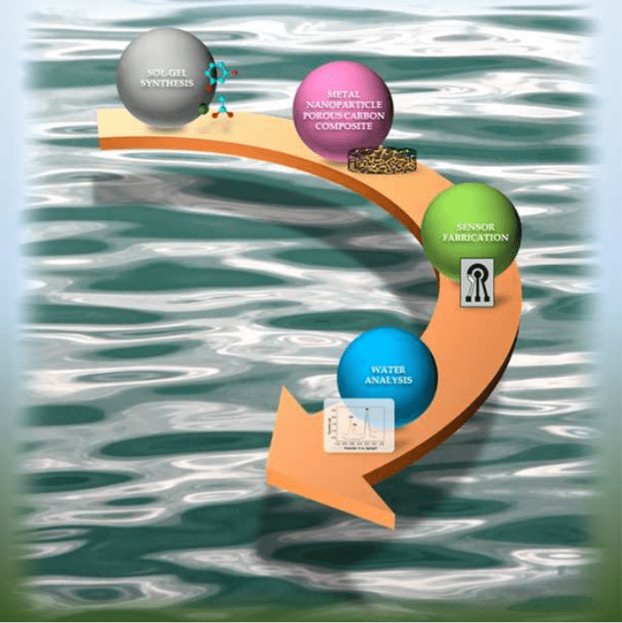
Congratulations to the authors Martí Gich, Anna Roig (N&N group), Pengfei Niu (former member of the N&N group) and César Fernández‐Sánchez (N&N collaborator form the Barcelona Institute of Microelectronics) for their published paper “Metal Nanoparticle Carbon Gel Composites in Environment in Water Sensing Applications”. The manuscript was published in the journal Chemical Record in a special issue dedicated to Prof. Ruiz-Hitzky.
Abstract: The synthesis of organic‐inorganic nanocomposites that can interact with different environmental pollutants and can be mass‐produced are very promising materials for the fabrication of chemical sensor devices. Among them, metal (or metal oxide) nanoparticles doped conductive porous carbon composites can be readily applied to the production of electrochemical sensors and show enhanced sensitivity for the measurement of water pollutants, thanks to the abundant accessible and functional sites provided by the interconnected porosity and the metallic nanoparticles, respectively. In this personal account, an overview of several synthesis routes of porous carbon composites containing metallic nanoparticles is given, paying special attention to those based on sol‐gel techniques. These are very powerful to synthesize hybrid porous materials that can be easily processed into powders and thin films, so that they can be implemented in electrode fabrication processes based on screen‐printing and lithography techniques, respectively. We emphasize the sol‐gel routes developed in our group for the synthesis of bismuth or gold nanoparticle doped porous carbon composites applied to fabricate electrochemical sensors that can be scaled down to produce miniaturized on‐chip sensing devices for the sensitive detection of heavy metal pollutants in water. The trend towards the miniaturization of electrochemical sensors to be readily employed as analytical tools in environmental monitoring follow the market requirements of rapid and accurate on‐site analysis, small sample consumption and waste production, as well as potential for continuous or semi‐continuous in‐situ determination of a wide variety of target analytes.

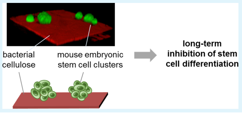
Congratulations to Anna Laromaine, Anna Roig and their collaborators from the Karlsruhe Institute of Technology Tina Tronser (first author) and Pavel A. Levkin for their new paper: Bacterial Cellulose Promotes Long-Term Stemness of mESC! This manuscript was published on ACS Applied Materials and Interfaces the 20th of April 2018.
Abstract: Stem cells possess unique properties, such as the ability to self-renew and the potential to differentiate into an organism’s various cell types. These make them highly valuable in regenerative medicine and tissue engineering. Their properties are precisely regulated in vivo through complex mechanisms that include multiple cues arising from the cell interaction with the surrounding extracellular matrix, neighboring cells, and soluble factors. Although much research effort has focused on developing systems and materials that mimic this complex microenvironment, the controlled regulation of differentiation and maintenance of stemness in vitro remains elusive. In this work, we demonstrate, for the first time, that the nanofibrous bacterial cellulose (BC) membrane derived from Komagataeibacter xylinus can inhibit the differentiation of mouse embryonic stem cells (mESC) under long-term conditions (17 days), improving their mouse embryonic fibroblast (MEF)-free cultivation in comparison to the MEF-supported conventional culture. The maintained cells’ pluripotency was confirmed by the mESCs’ ability to differentiate into the three germ layers (endo-, meso-, and ectoderm) after having been cultured on the BC membrane for 6 days. In addition, the culturing of mESCs on flexible, free-standing BC membranes enables the quick and facile manipulation and transfer of stem cells between culture dishes, both of which significantly facilitate the use of stem cells in routine culture and various applications. To investigate the influence of the structural and topographical properties of the cellulose on stem cell differentiation, we used the cellulose membranes differing in membrane thickness, porosity, and surface roughness. This work identifies bacterial cellulose as a novel convenient and flexible membrane material enabling long-term maintenance of mESCs’ stemness and significantly facilitating the handling and culturing of stem cells.

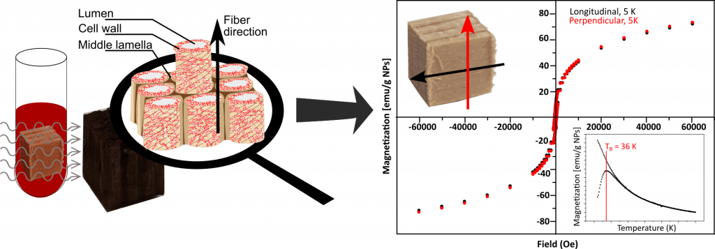
Congratulations to the authors: Jana S. Segmehl, Anna Laromaine, Tobias Keplinger, Anna May-Masnou, Ingo Burgertab and Anna Roig for their new publication entitled: Magnetic wood by in situ synthesis of iron oxide nanoparticles via a microwave-assisted route. The paper was first published on 9th of March 2018 in the Journal of Materials Chemistry.
Abstract:
Functional materials with high porosity and hierarchical structures are highly demanded for numerous material applications. In this study a magnetic hybrid material derived from wood and superparamagnetic iron oxide nanoparticles (SPIONs), was synthesized by microwave-assisted thermal decomposition. This novel in situfunctionalization approach resulted in a homogeneous distribution of the integrated inorganic component within the entire complex wood cell wall structure, which was previously not achieved. In a detailed investigation based on confocal Raman microscopy imaging, transmission electron microscopy, and optical microscopy the precipitated phase and the resulting hybrid structure were characterized. Magnetic measurements revealed the impact of the anisotropic wood scaffold on the integrated magnetic functionality and confirmed the isotropic superparamagnetic characteristics of the in situ precipitated nanoparticles. Therewith, it is clearly demonstrated, that the anisotropic properties of the obtained hybrid material result from the particle organization in the given spruce wood structure and no alteration of particle properties is induced by the presence of the lignocellulosic material.

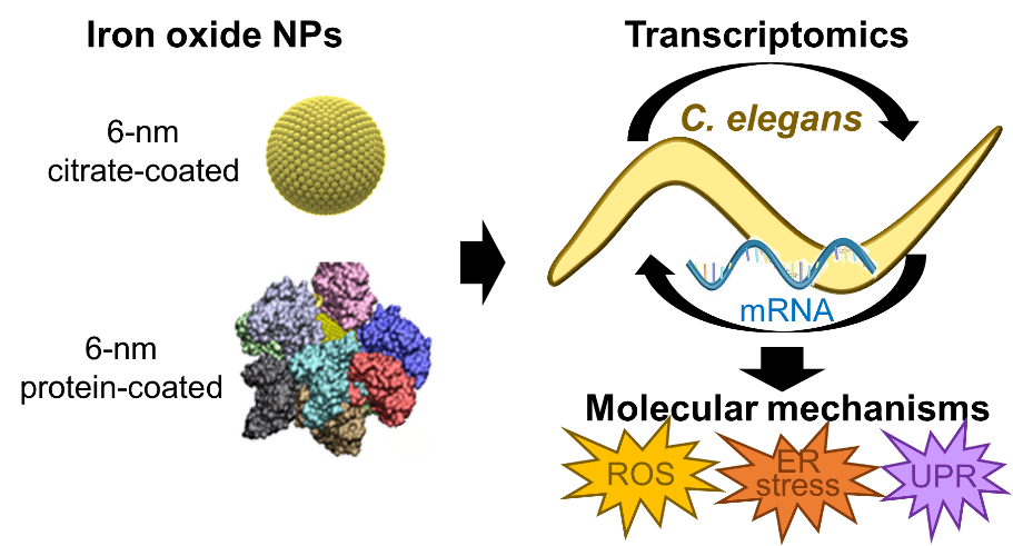 We congratulate the autos Laura Gonzalez-Moragas, Si-Ming Yu, Núria Benseny-Cases, Stephen Stürzenbaum, Anna Roig and Anna Laromaine for their recently accepted paper: Toxicogenomics of iron oxide nanoparticles in the nematode C. elegans. This research arises from a collaboration of the N&N group, the ALBA Synchrotron Light Source (MIRAS line) and the King’s College London.
Here you can read the abstract of the paper:
We present a mechanistic study of the effect of iron oxide nanoparticles (SPIONs) in C. elegans combining a genome-wide analysis with the investigation of specific molecular markers frequently linked to nanotoxicity. The effects of two different coatings were explored: citrate, an anionic stabilizer, and bovine serum albumin, as a pre-formed protein corona. The transcriptomic study identified differentially expressed genes following an exposure to SPIONs. The expression of genes involved in oxidative stress, metal detoxification response, endocytosis, intestinal integrity and iron homeostasis was quantitatively evaluated. The role of oxidative stress was confirmed by gene expression analysis and by synchrotron Fourier Transform infrared microscopy based on the higher tissue oxidation of NP-treated animals. The observed transcriptional modulation of key signaling pathways such as MAPK and Wnt suggests that SPIONs might be endocytosed by clathrin-mediated processes, a putative mechanism of nanotoxicity which deserves further mechanistic investigations.
Citation:
Toxicogenomics of iron oxide nanoparticles in the nematode C. elegans
Laura Gonzalez-Moragas, Si-Ming Yu, Núria Benseny-Cases, Stephen Stürzenbaum, Anna Roig and Anna Laromaine
Nanotoxicology, Accepted manuscript online: 15 Jun 2017. DOI: 10.1080/17435390.2017.1342011

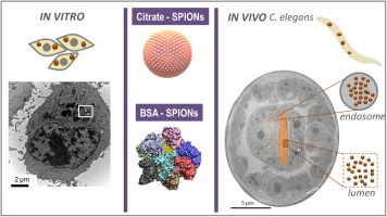 The article “Bio-identity and fate of albumin-coated SPIONs evaluated in cells and by the C. elegans model” (Si-Ming Yu, Laura González-Moragas, Maria Milla, Androniki Kolovou, Rachel Santarella-Mellwig, Yannick Schwab, Anna Laromaine, Anna Roig) is now published online in Acta Biomaterialia at:
http://www.sciencedirect.com/science/article/pii/S174270611630349X.
doi:10.1016/j.actbio.2016.07.024.
Abstract: Nanoparticles, whose surface adsorbs proteins in an uncontrolled and non-reproducible manner will have limited uses as nanomedicinal products. A promising approach to avoid nanoparticle non-specific interactions with proteins is to design bio-hybrids by purposely pre-forming a protein corona around the inorganic cores. Here, we investigate, in vitro and in vivo, the newly acquired bio-identity of superparamagnetic iron oxide nanoparticles (SPIONs) upon their functionalization with a pre-formed and well-defined bovine serum albumin (BSA) corona. Cellular uptake, intracellular particle distribution and cytotoxicity were studied in two cell lines: adherent and non-adherent cells. BSA decreases nanoparticle internalization in both cell lines and protects the iron core once they have been internalized. The physiological response to the nanoparticles is then in vivo evaluated by oral administration to Caenorhabditis elegans, which was selected as a model of a functional intestinal barrier. Nanoparticle biodistribution, at single particle resolution, is studied by transmission electron microscopy. The analysis reveals that the acidic intestinal environment partially digests uncoated SPIONs but does not affect BSA-coated ones. It also discloses that some particles could enter the nematode’s enterocytes, likely by endocytosis which is a different pathway than the one described for the worm nutrients.
Keywords: Iron oxide nanoparticles; Protein corona; Cytotoxicity; C. elegans; Biodistribution.
Congratulations to all the authors!

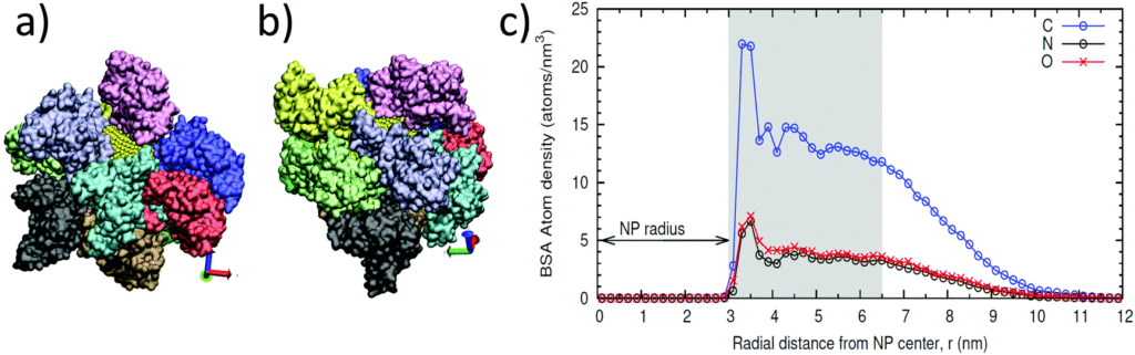
Albumin-coated SPIONs: An experimental and theoretical evaluation of protein conformation, binding affinity, and competition with serum proteins, by Siming Yu, Alex Perálvarez-Marín, Caterina Minelli, Jordi Faraudo, Anna Roig* and Anna Laromaine*, has just been accepted for publication in Nanoscale, DOI: 10.1039/C6NR01732K.
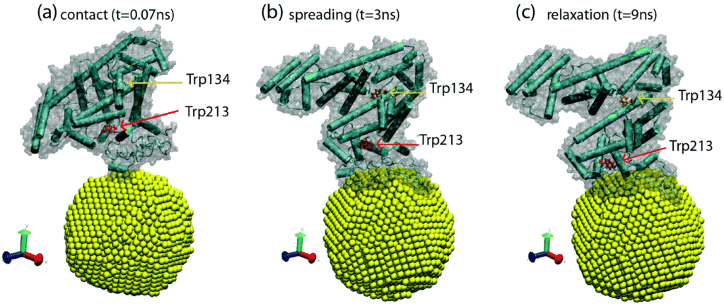 Among inorganic nanoparticles, superparamagnetic iron oxide nanoparticles (SPIONs) show great promise for medicine. In this work, we study in detail the formation, composition, and structure of a monolayer of bovine serum albumin (BSA) on SPIONs. We determine, both by molecular simulations and experimentally, that ten molecules of BSA form a monolayer of BSA around the SPIONs and their binding strength to the SPIONs is about 3.5×10⁻⁴ M, ten times higher than the adsorption of fetal bovine serum (FBS) on the same SPIONs. We elucidate a strong electrostatic interaction between BSA and the SPIONs, although the secondary structure of the protein is not affected. We present data that supports the strong binding of the BSA layer on SPIONs and the properties of the BSA layer as a protein-resistant coating. We believe that a complete understanding of the behavior and morphology of BSA-SPIONs and how the protein interacts with SPIONs is crucial for improving NP surface design and expanding the potential applications of SPIONs in nanomedicine.

Today is the big day: second manuscript accepted!
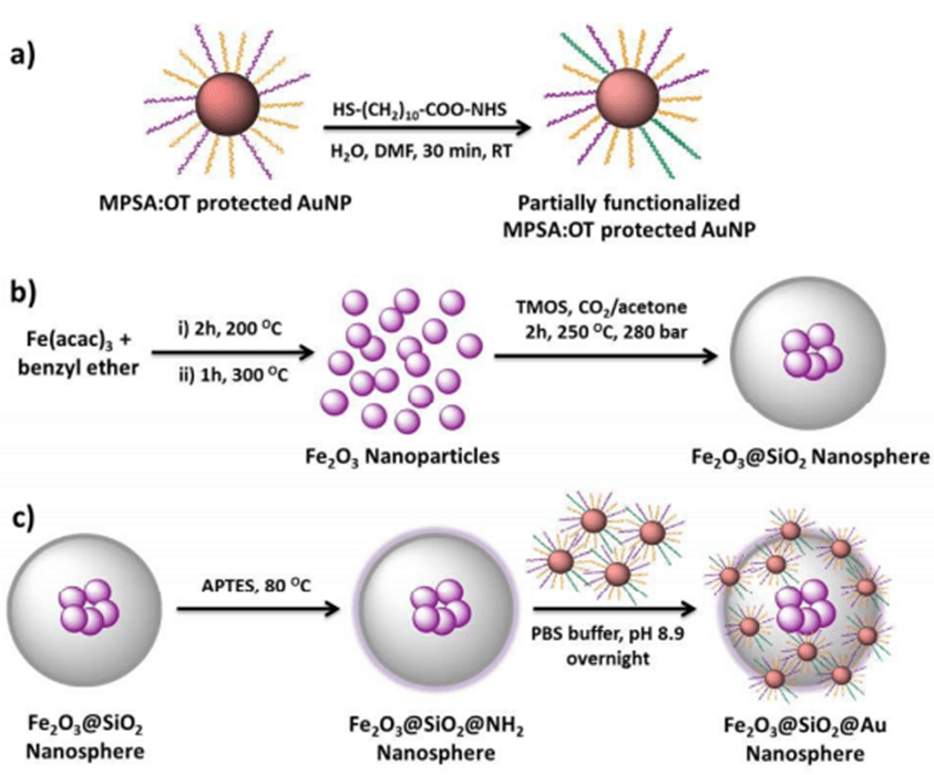
The manuscript “A silica-based magnetic platform decorated with mixed ligand gold nanoparticles: A recyclable catalyst for esterification reactions” (Elif Ertem, Nerea Murillo-Cremaes, Randy Patrick Carney, Anna Laromaine, Emma-Rose Janeček, Anna Roig* and Francesco Stellaccia*; DOI: 10.1039/C6CC01146B) has been accepted in Chemical Communications.
This paper describes a novel and convenient synthetic strategy for the preparation of magnetically responsive silica nanospheres decorated with mixed ligand protected gold nanoparticles. Gold nanoparticles are attached to the silica surface via stable amide bond formation. The hierarchical nanospheres show promising results as a reusable and efficient catalyst for esterification reactions and they can be recovered through a simple magnetic separation.
Congratulations!
Figure: Schematic illustration of: (a) the partial ligand exchange of 3-mercapto-1-propanesulfonate (MPSA):1-octanethiol(OT) covered gold nanoparticles, (b) the synthesis of core-shell magnetic silica and (c) the synthesis of the magnetic silica decorated with gold nanoparticles hierarchical nanospheres.

 We are very happy to announce that the manuscript “Gold nanotriangles decorated with superparamagnetic iron oxide nanoparticles: a compositional and microstructural study“ (J.A. Hachtela, S. Yuc , A.R. Lupini, S.T. Pantelidesa, M. Gich, A. Laromaine, A. Roig, DOI: 10.1039/C6FD00028B) has been accepted by the Faraday Discussions journal.
The manuscript will be presented and discussed at a forthcoming Faraday Discussions meeting, this next summer. During the meeting, all delegates will be able to contribute to the debate, which will be included in the final volume. This will be a great opportunity to discuss the formation mechanism of the magnetic gold nanotriangles with some experts on this field.
The combination of iron oxide and gold in a single nanoparticle results in both magnetic and plasmonic properties that can stimulate novel applications in bio-sensing, medical imaging, or therapeutics. Microwave heating method allows the fabrication of multi-component, multi-functional nanostructures by promoting selective heating at desired sites. Recently, we reported a microwave-assisted polyol route yielding gold nanotriangles decorated with iron oxide nanoparticles (1). Here, we present an in-depth microstructural and compositional characterization of the system by using scanning transmission electron microscopy (STEM) and electron energy loss (EELS) spectroscopy. A method to remove the iron oxide nanoparticles from the gold nanocrystals and some insights on crystal nucleation and growth mechanisms are also provided.
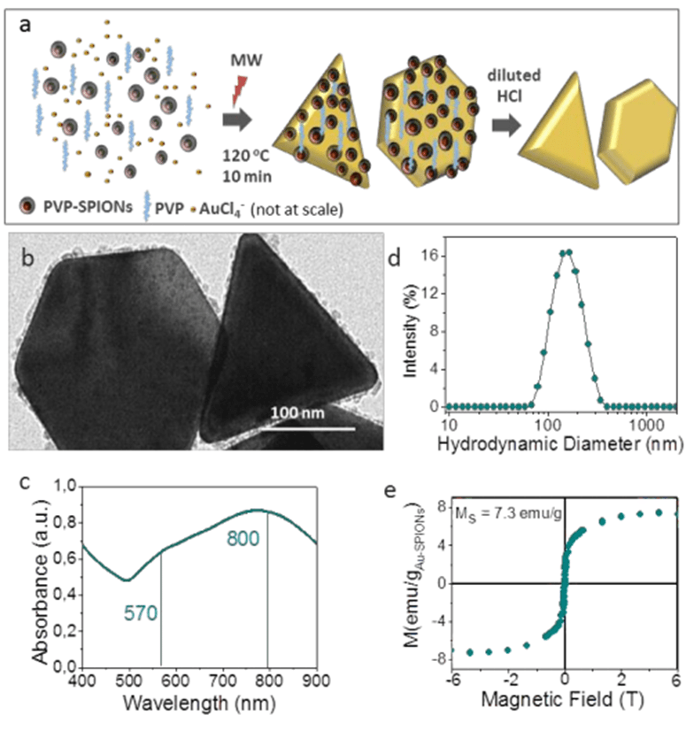
Figure: (a) Schematic representation of the synthesis route. (b) HRTEM image of a Au NH-SPIONs and a Au NT-SPIONs. Characterization of the heterostructures: (c) UV-Vis-NIR spectra. (d) Hydrodynamic diameter of the Au-SPIONS measured by DLS. (e) Magnetization curve up to 6 T at 5K.
(1) Magnetic gold nanotriangles by microwave polyol synthesis (S.M. Yu, J.A. Hachtel, M.F. Chisholm, S.T. Pantelides, A. Laromaine, A. Roig; Nanoscale 2015, 7, 14039-14046. DOI: 10.1039/C5NR03113C).

The work has resulted from a collaboration between the ICMAB, the CNM and the company Dropsens.
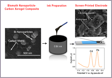
Screen-printed electrodes made of a bismuth nanoparticle porous carbon nanocomposite material applied to the detection of heavy metals (Pengfei Niu, César Fernández-Sánchez,* Martí Gich,* Carla Navarro-Hernández, Pablo Fanjul-Bolado, and Anna Roig, Microchimica Acta, , Volume 183, Issue 2, pp 617-623).
This work reports on the simplified fabrication and on the characterization of bismuth-based screen-printed electrodes (SPEs) for use in heavy metal detection.
A nanocomposite consisting of bismuth nanoparticles and amorphous carbon was synthesized by a combined one-step sol-gel and pyrolysis process and milled down to a specific particle size distribution as required for the preparation of an ink formulation to be used in screen printing. The resulting electrochemical devices were applied to the detection of Pb(II) and Cd(II) ions in water samples.
The porous structure of carbon and the high surface area of the bismuth nanoparticles allow for the detection of Pb(II) and Cd(II) at concentration levels below 4 ppb. The application of the SPEs was demonstrated by quantifying these ions in tap drinking water and wastewater collected from an influent of an urban wastewater treatment plant.
The N&N group participates in the @NextGEM_eu project, for which we prepared this video explaining how we work with c.elegans. 🪱
👉 Dr. @PolAlonso1 explains how we use these nematodes and why they are useful for assessing biological effects.
#BioEM #5G
We are thrilled to announce that Dr. @AnnaRoig8 from the @NNgroupICMAB research group (@icmabCSIC, Barcelona, Spain) will be offering a talk on "Opportunities of bacterial nanocellulose in healthcare". This Wednesday at 12.15h at the BBT Weekly Meetings, at @EEBE_UPC