
Hot off the press: New paper in Acta Biomaterialia!
New paper “Sciatic nerve regeneration after traumatic injury using magnetic targeted adipose-derived mesenchymal stem cells“ has been published in the journal Acta Biomaterialia.

New paper “Sciatic nerve regeneration after traumatic injury using magnetic targeted adipose-derived mesenchymal stem cells“ has been published in the journal Acta Biomaterialia.

 The paper “In vivo testing of gold nanoparticles using the Caenorhabditis elegans model organism” has been publised in Acta Biomaterialia (Available online 1 February 2017; doi: 10.1016/j.actbio.2017.01.080).
The authors of the paper are Laura González-Moragas, Pascal Berto, Clara Vilches, Romain Quidant, Androniki Kolovou, Rachel Santarella-Mellwig, Yannick Schwab, Stephen Stürzenbaum, Anna Roig, and Anna Laromaine.
The paper is a result of a collaboration between the NN Group at ICMAB (González-Moragas, Roig and Laromaine), the ICFO-Institut de Ciències Fotòniques (Berto, Vilches and Quidant), the European Molecular Biology Laboratory, EMBL in Heidelberg, Germany (Kolovou, Santarella-Mellwig, Schwab) and King’s College London in UK (Stürzenbaum).
Congratulations!
Abstract:
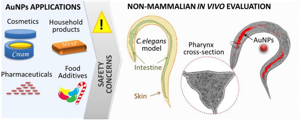
Gold nanoparticles (AuNPs) are present in many man-made products and cosmetics, and are also used by the food and medical industries. Tight regulations regarding the use of mammalian animals for product testing can hamper the study of the specific interactions between engineered nanoparticles and biological systems. Invertebrate models, such as the nematode Caenorhabditis elegans (C. elegans), can offer alternative approaches during the early phases of nanoparticle discovery.
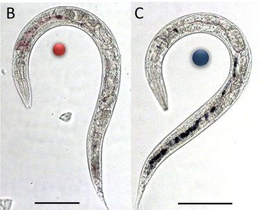
Here, we thoroughly evaluated the biodistribution of 11-nm and 150-nm citrate-capped AuNPs in the model organism C. elegans at multiple scales, moving from micrometric to nanometric resolution and from the organism to cellular level. We confirmed that the nanoparticles were not able to cross the intestinal and dermal barriers. We investigated the effect of AuNPs on the survival and reproductive performance of C. elegans, and correlated these effects with the uptake of AuNPs in terms of their number, surface area, and metal mass. In general, exposure to 11-nm AuNPs resulted in a higher toxicity than the larger 150-nm AuNPs. NP aggregation inside C. elegans was determined using absorbance microspectroscopy, which allowed the plasmonic properties of AuNPs to be correlated with their confinement inside the intestinal lumen, where anatomical traits, acidic pH and the presence of biomolecules play an essential role on NP aggregation. Finally, quantitative PCR of selected molecular markers indicated that exposure to AuNPs did not significantly affect endocytosis and intestinal barrier integrity.

 The article “Bio-identity and fate of albumin-coated SPIONs evaluated in cells and by the C. elegans model” (Si-Ming Yu, Laura González-Moragas, Maria Milla, Androniki Kolovou, Rachel Santarella-Mellwig, Yannick Schwab, Anna Laromaine, Anna Roig) is now published online in Acta Biomaterialia at:
http://www.sciencedirect.com/science/article/pii/S174270611630349X.
doi:10.1016/j.actbio.2016.07.024.
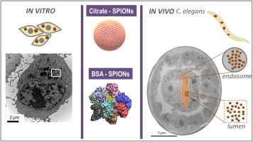
Abstract: Nanoparticles, whose surface adsorbs proteins in an uncontrolled and non-reproducible manner will have limited uses as nanomedicinal products. A promising approach to avoid nanoparticle non-specific interactions with proteins is to design bio-hybrids by purposely pre-forming a protein corona around the inorganic cores. Here, we investigate, in vitro and in vivo, the newly acquired bio-identity of superparamagnetic iron oxide nanoparticles (SPIONs) upon their functionalization with a pre-formed and well-defined bovine serum albumin (BSA) corona. Cellular uptake, intracellular particle distribution and cytotoxicity were studied in two cell lines: adherent and non-adherent cells. BSA decreases nanoparticle internalization in both cell lines and protects the iron core once they have been internalized. The physiological response to the nanoparticles is then in vivo evaluated by oral administration to Caenorhabditis elegans, which was selected as a model of a functional intestinal barrier. Nanoparticle biodistribution, at single particle resolution, is studied by transmission electron microscopy. The analysis reveals that the acidic intestinal environment partially digests uncoated SPIONs but does not affect BSA-coated ones. It also discloses that some particles could enter the nematode’s enterocytes, likely by endocytosis which is a different pathway than the one described for the worm nutrients.
Keywords: Iron oxide nanoparticles; Protein corona; Cytotoxicity; C. elegans; Biodistribution.
Congratulations to all the authors!
The N&N group participates in the @NextGEM_eu project, for which we prepared this video explaining how we work with c.elegans. 🪱
👉 Dr. @PolAlonso1 explains how we use these nematodes and why they are useful for assessing biological effects.
#BioEM #5G
We are thrilled to announce that Dr. @AnnaRoig8 from the @NNgroupICMAB research group (@icmabCSIC, Barcelona, Spain) will be offering a talk on "Opportunities of bacterial nanocellulose in healthcare". This Wednesday at 12.15h at the BBT Weekly Meetings, at @EEBE_UPC